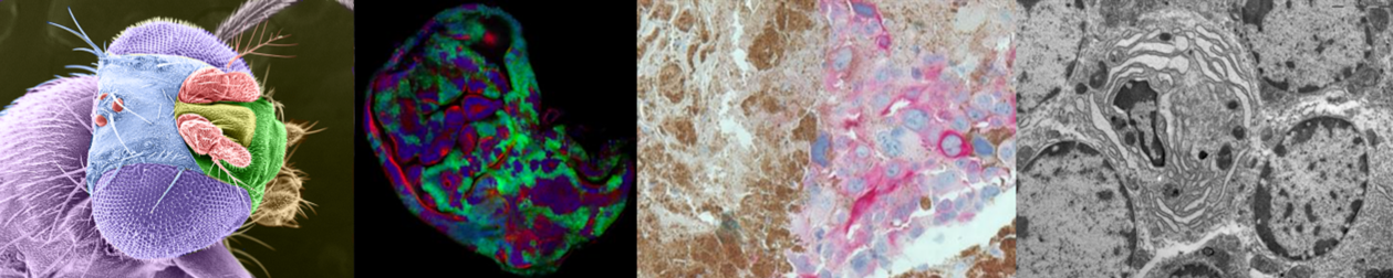
Peter MacCallum’s Centre for Advanced Histology and Microscopy (CAHM) underpins a multitude of cancer research projects and houses four core platforms:
1. Histology: inclusive of wax and cryo-sections, special stains, and a comprehensive immunohistochemistry suite.
2. Optical Microscopy: houses high-end optical microscopes including laser scanning confocal microscopes, spinning disk confocal, multi photon microscope (usable for intra-vital imaging), slide scanner, structured illumination microscopy, and single molecule localisation microscopy.
3. Spatial Biology: inclusive of training and optimisation of mIHC projects, and equipment including auto-stainers, imaging equipment, and other spatial-biology techniques
3. Image Analysis: Sophisticated software for analysing images.
Importantly, researchers utilising the facility receive support, training, and advice from expert technical scientists.
At CAHM, sustainability is a top priority! We're committed to minimizing our environmental impact and are actively pursuing LEAF certification across our platforms. We invite all researchers to join us by adopting sustainable habits in your research. Together, we can make a real difference and build a greener future for science!
Want to learn more? Get in touch with Greenlabs@petermac.org for details on the LEAF program and Green Labs initiatives.
The Peter MacCallum Advanced Histology Platform provides a histology service dedicated to research.
Services and Support Offered
Key Equipment

The Peter MacCallum Advanced Optical Microscopy Platform incorporates multiple imaging modalities including wide-field microscopy, laser capture microscopy, laser scanning confocal microscopy, multi-photon intravital microscopy, and super resolution microscopy. The multi-photon microscope is a recent addition and was purchased alongside the Zeiss Elrya super resolution confocal microscope (TIRF, SIM, PALM, dSTORM) and the fully automated slide scanner through a successful $3.5m grant from the Australian Cancer Research Foundation (ACRF). Five microscopes are dedicated to live cell imaging and are equipped with heated environmental chambers, gas control and motorized stages.
Services and Support Offered
Key Equipment
Confocal Laser Scanning Microscopes
Spinning Disk Confocal Microscope
Super Resolution Microscope
Multiphoton Microscope
Light Sheet Microscope
Widefield Microscopes (all equipped with DIC and epi-fluorescence)
Automated slide scanner
Spatial Proteomics & Transcriptomics analysis

The Image Analysis Platform provides a number of high-end image analysis software programs for powerful multi-dimensional analysis. Software is also provided to design custom items for specialist imaging requirements using our 3D printer.
Key Equipment
|
Ilia Voskoboinik (Chair) |
Louise Cheng (Deputy-Chair) |
Julie Blasioli |
Metta Jana |
|
Andrew Cox |
Vi Wickramasinghe |
Katie Owen |
Kazuhide Okuda |
|
Marcos Sande-Melon |
Jesse Rudd-Schmidt |
Richard Young |
Dani Tatuka |
|
Hugo Saunders |
Thu Nguyen |
Josh Hudson |
|
Nicholas Anthony - Manager Centre for Advanced Histology and Microscopy
Metta Jana - Deputy Manager Centre for Advanced Histology and Microscopy
Use of the facility is available to external users, subject to availability.
Contact: Nicholas Anthony (nicholas.anthony@petermac.org, 03 8559 7569) for enquiries relating to the use of facilities at CAHM and project design/advice.
| Hours | Location |
|
Available to trained users 24/7 |
Level 8 |
PMCC Researchers, please head to our Connect webpage for user guides, educational resources, and up-to-date information.
External Researchers, can view our public website for information.
Head: Nicholas Anthony
Core staffs: Metta Jana, Judy Borg, Han Aw Yeang, Dhanya Menon, Basia Przybylowski, Rejhan Idrizi, Ashley Rozario
| Name | Role | Phone | Location | |
|---|---|---|---|---|
| Nicholas Anthony |
Manager, Centre for Advanced Histology and Microscopy
|
85597569
|
nicholas.anthony@petermac.org
|
Level 8
|
| Service list |
| ► Histology - Additional Services (1) | |||
| Name | Description | Price | |
|---|---|---|---|
| Histology - Training on Leica autostainer XL and Dako coverslipper |
Training on use of Leica autostainer XL and Dako coverslipper |
Inquire | |